Has the mystery of tardigrades’ extreme resilience finally been solved?
September 2007. A Russian rocket lifts off from the Baikonur Cosmodrome in Kazakhstan, carrying strange little creatures just one millimeter long on a ten-day journey around the Earth. These are tardigrades participating in the European Space Agency’s (ESA) Foton-M3 space program.
Simon Galas, University of Montpellier and Myriam Ricard, University of Montpellier
It’s a curious twist of fate for these astronaut tardigrades, which were all aquatic when they first appeared on Earth (probably during the Cambrian period, about 500 million years ago).
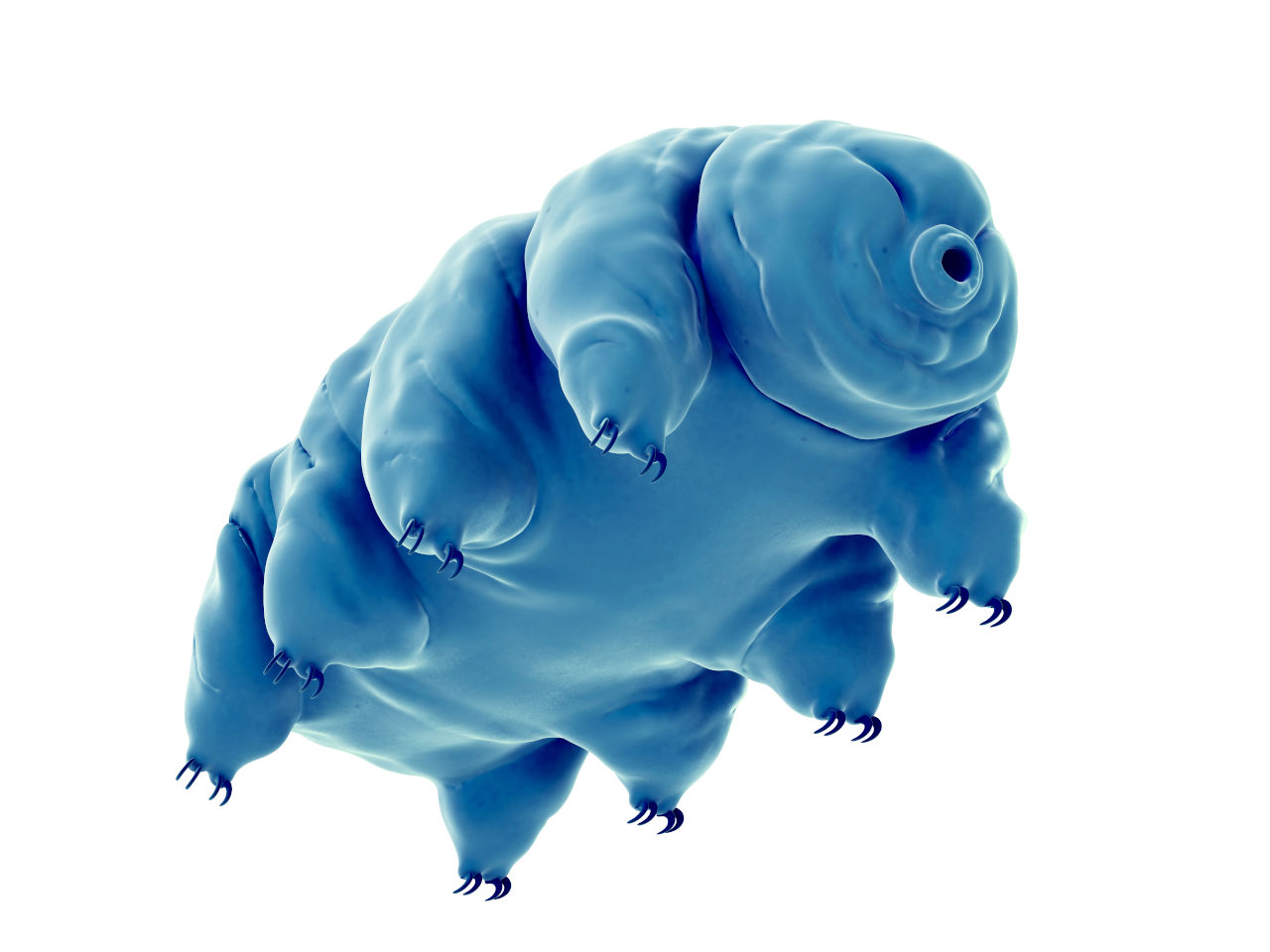
Tardigrades, also known as water bears, have four pairs of legs and a pair of small eyes. With 1,400 known species today, they can be found all over the Earth, from the peaks of the Himalayas to the depths of the oceans.
Tardigrades can lose up to 95% of their water content
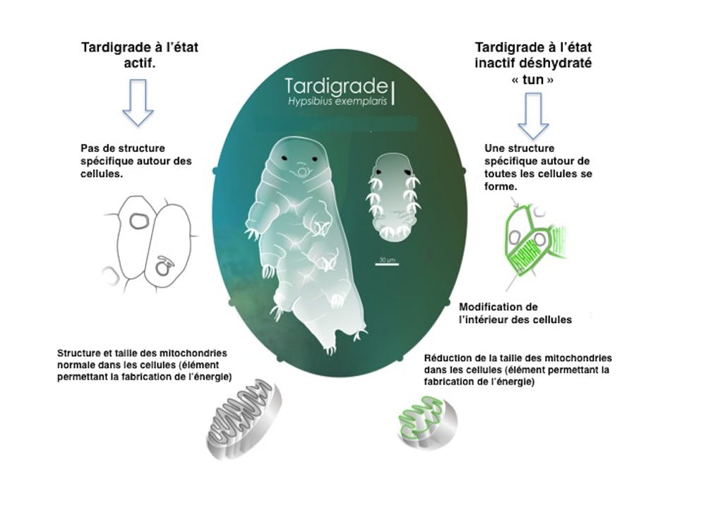
Terrestrial tardigrade species retain a memory of their aquatic origins. As soon as the thin film of water covering its body disappears, the tardigrade undergoes an active transformation that causes it to lose 95% of its water content and shrink in size by nearly 40%. In this state, the tardigrade resembles a small barrel, hence the name “tun” used by English speakers to refer to it in this particular state, which is a state ofanhydrobiosis.
Anhydrobiosis is a state of suspended animation caused by dehydration. In this unique state, tardigrades are able to withstand extreme conditions.
During this mission in low Earth orbit (at an altitude of 258–281 km above sea level), these tardigrades in a state of suspended animation were exposed to the combined effects of the vacuum of space, cosmic rays, and extreme ultraviolet radiation.
Once back on Earth, 12% of these astronaut tardigrades were successfully revived simply by placing a drop of water on their bodies. This spectacular revival, which takes less than five minutes in some species, astonished scientists and established tardigrades as the only animals capable of surviving the combined effects of the vacuum of space, cosmic radiation, and ultraviolet rays in real-world conditions.
One might think that this state of anhydrobiotic dormancy, or “tune,” allows tardigrades to withstand only the vacuum of space and its low pressure ( 10⁻⁶ pascals), but that is not the case. Tardigrades in the same anhydrobiotic state have been subjected to pressures of up to 7.5 gigapascals. This pressure is equivalent to the weight of the rock pressing down on your shoulders if you were to descend 180 km into the Earth’s mantle!
What happens inside the cells of dehydrated tardigrades?
Until now, no one had tried to find out what happens inside a tardigrade when it turns into a "tun."
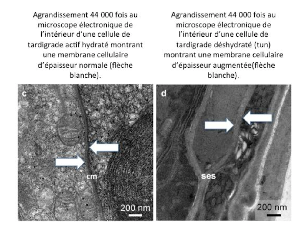
By bringing together two laboratories from the CNRS and the University of Montpellier, we were able to produce images using high-resolution microscopy techniques for the first time. The images captured using transmission electron microscopy allowed us to take a journey inside a tardigrade anhydrobiote.
An electron microscope uses electrons to illuminate the object being observed and can magnify it up to 5 million times, whereas an optical microscope, which illuminates the object with light (photons), can magnify it by a maximum of 2,000 times. Electron microscopes are capable, for example, of taking pictures of viruses.
The observation of these images of tardigrades was the subject of a scientific paper published in the international journal Nature Scientific Reports.
Simon Galas, Author provided
It all begins with the animal’s dehydration, which results in the loss of nearly all its water. At the same time, the interior of the tardigrade reorganizes itself, and all the structures that make up a tardigrade cell remain unchanged—for example, the cell nucleus, which contains the chromosomes, and the mitochondria, which produce the cell’s energy. In fact, the entire cell structure appears as a miniaturized version of the original. The cells are reduced in size by nearly 40%.
Something unexpected appears in the images
A molecular barrier surrounding all of the tardigrade’s cells gradually forms, reaching a thickness of up to 100 nanometers in some places. This is extremely thick compared to the membrane that normally surrounds a cell, even in humans. This intercellular barrier, unknown until now, may be the secret behind the tardigrade’s resilience, allowing it to withstand extreme pressures without being pulverized—pressures that no other living organism could survive. Research into this barrier could potentially lead to the discovery of new ultra-resistant materials for a future industry that is both innovative and environmentally friendly.
Simon Galas, Author provided
One theory may help explain what this unique protective barrier is made of. Professor Takekazu Kunieda of the University of Tokyo recently discovered a specific class of proteins in tardigrades that are capable of vitrifying dehydrated tardigrades, as demonstrated by a laboratory at the University of North Carolina in the United States.
But what happens to this intercellular barrier when a drop of water is placed on the back of an anhydrobiotic tardigrade? Electron microscope images show that the barrier gradually disappears as the tardigrade emerges from its state of suspended animation, vanishing completely after just 24 hours.
We do not yet know whether this newly discovered protective barrier is produced only by the species of tardigrade (Hypsibius exemplaris) raised in our laboratory or by other species of tardigrades as well.
Recent sequencing data from the first tardigrade genomes have revealed some surprises, such as the presence of a set of genes unknown in other living species, whose functions biologists are only just beginning to study. This discovery sheds even more light on tardigrades’ ability to protect themselves against all kinds of threats and to achieve the high levels of resilience for which they are renowned.![]()
Simon Galas, Professor of Genetics and Molecular Biology of Aging, CNRS – School of Pharmacy, University of Montpellier and Myriam Ricard, PhD in Molecular and Cellular Biology, University of Montpellier
This article is republished from The Conversation under a Creative Commons license. Readthe original article.