Chemical pollution and the cocktail effect: a path toward toxicological testing without animal experimentation
Here is an image showing mitochondria in pink: these are the lungs and “powerhouses” that enable cells (shown in green, with their nuclei in blue) to breathe, live, and perform their functions.
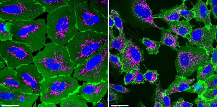
Abdel Aouacheria, University of Montpellier
In the healthy cells on the left, the mitochondria are relatively long and interconnected, resembling a road network viewed from above; whereas in the stressed and damaged cells on the right, their network has broken down into a constellation of isolated mitochondria, which produce less energy and will eventually drive the cells toward cell death.
Thus, mitochondria serve as a good indicator of our cells’ health, with the architecture of their networks even varying in tissues from sick individuals. Thanks to real-timeconfocal imaging, particularly using high-content imaging robots, we can reveal in less than a second the contours of a living cell, its nucleus, and unique “organelles” (the components of a cell that perform specific functions, such as mitochondria), in order to study the effects of various pollutants on cells and their health.
Rapid mitochondrial imaging as an “early warning system” in environmental health
Air, water, and food contamination, soil pollution, and noise pollution: measuring the impact of environmental risks on the health of organisms and ecosystems is no easy task.
The images generated by imaging platforms are processed by computers to provide valuable insights into the effects of the many pollutants that surround us.
This marks a step forward in understanding the “exposome,” a concept introduced by the British scientist Christopher Paul Wild in 2005 and defined as the totality of exposures to which an individual is subjected throughout their life (from conception to death).
In fact, outdoor pollution (air, water, soil) and indoor pollution (homes, offices, cars) not only have negative effects on our health (by triggering chronic diseases) and on ecosystems (which are experiencing an unprecedented decline in biodiversity and agricultural yields), but also entail enormous socioeconomic costs.
It is estimated to be responsible for one in six deaths each year— three times more than AIDS, tuberculosis, and malaria combined. We are also constantly living in a “chemosphere” (a mixture of substances) whose risks are poorly understood: epidemiologists and toxicologists have been able to assess the toxicity of only a tiny fraction of the 350,000 chemicals registered since the 1960s in the major national and regional chemical inventories (for production and commercial use).
The health and environmental impacts of these compounds, which could jeopardize the integrity of the Earth system, remain poorly understood when studied in isolation. Furthermore, their potential “cocktail effects” (effects resulting from simultaneous exposure to multiple substances, which are sometimes more harmful than exposure to a single substance) are almost never tested due to a lack of appropriate technologies.
How can we capture the reality of these multiple exposures at the cellular level within an organism?
A new generation of toxicology tests that do not involve animal testing
This is where mitochondria come into play. The fragmentation of mitochondria and the mitochondrial network is, in fact, an early indicator of their loss of function and thus a marker of environmental stress.
The method involves staining (with vital dyes) the mitochondria and other components of cells cultured in vitro. These cells are primary human cells or cell lines derived from various tissue sources (such as skin, lung, kidney, and intestine) and are exposed to toxic substances found in our environment, such as pesticides or fine particulate matter. There is no need here to sacrifice an animal for each experiment.
Using specialized software, a set of parameters is calculated from confocal microscopy images: mitochondrial size, circularity, and connectivity are among the hundred or so possible descriptors that help identify the harmful effects of toxic substances, either alone or in combination (cocktail effect).
The goal is to link initiating molecular events, such as exposure to these chemicals, with harm or toxicity across various biological scales—from cells and tissues to organs and individuals. While mitochondria link toxicities and alterations occurring at the microscopic level (cells) to the adverse effects and pathologies observed at the tissue level, uncertainty remains regarding the extrapolation of toxicological data obtained in vitro to potential effects on organisms and ecosystems.
Ultimately, this system could also help identify ingredients capable of protecting or restoring the “mitochondriomes” (the totality of mitochondria) in cells exposed to pollutants.
Abdel Aouacheria, biologist, research fellow at the CNRS, specialist in cell life and death, University of Montpellier
This article is republished from The Conversation under a Creative Commons license. Readthe original article.