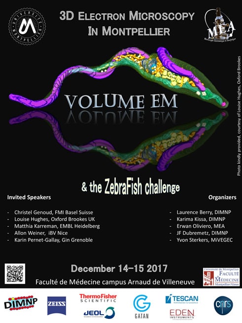
Conference “Volume EM – 3D Electron Microscopy”
This event has already taken place!
December 14 and 15, 2017, starting at 1:30 p.m.
New School of Medicine, Arnaud de Villeneuve Campus
 The Electron Microscopy and Analytics Platform and DIMNP are pleased to announce the “Volume EM” conference, which will be held on December 14 and 15 at the new medical school.
“Volume EM” refers to a set of revolutionary methods that enable very high-resolution 3D imaging of the ultrastructure of large sample volumes using scanning electron microscopes coupled with various automated serial sectioning techniques. These methods make it possible to obtain highly informative results in a matter of hours or days—results that would previously have required, at best, months or even years of work. Although still in its early stages in France, this type of approach is experiencing rapid growth internationally and is finding applications in numerous fields of research.
The conference brings together leading scientists and development experts who will present the principles behind various methods and selected examples of their applications.
A “zebrafish challenge” was also presented to the manufacturers, asking them to image a specified section of a zebrafish embryo using the equipment of their choice. The goal was to test the hardware’s performance under real-world research conditions. The results will be presented and discussed at the close of the conference and will highlight the advantages and limitations of each type of method.
Registration is free; you can find all the information and the registration form here.
If you have any questions, pleasesend an email.
The University’s Electron Microscopy platform and DIMNP are pleased to announce the Volume EM conference, which will take place at the new “Fac de Médecine” (near the Occitanie tram station) on December 14–15.
Volume EM is a set of methods that enable the acquisition of high-resolution 3D ultrastructural data using scanning electron microscopy coupled with various automated serial sectioning techniques. With these methods, it is now possible to obtain high-resolution 3D data within a few hours or days. Volume EM is rapidly gaining international attention, with applications in many fields of research.
During the conference, leading scientists and application developers will present the principles of these methods and selected examples of their applications in research.
A“zebrafish challenge” has also been proposed to various microscope manufacturers, in which they were tasked with imaging a specified section of a zebrafish embryo using the equipment of their choice. The objective will be to analyze the results obtained under conditions that closely resemble “real research.” The data will be presented and discussed at the end of the conference, highlighting the advantages and limitations of each type of method.
The conference will be conducted entirely in English. Registration is free; you can find more information and fill out the registration form here.
If you have any questions, please send an email.
News

Decline of the Clam: The University of Montpellier, Ifremer, and Two Artists Launch the CLAM-EU Project to Engage Lagoon Communities Through Art and Science
Published on: July 20 , 2026
The University of Montpellier, Ifremer, painter Françoise Dagorn, and the pianist…